
Advantages and Applications of 3D Cell Microscope
2024-07-31
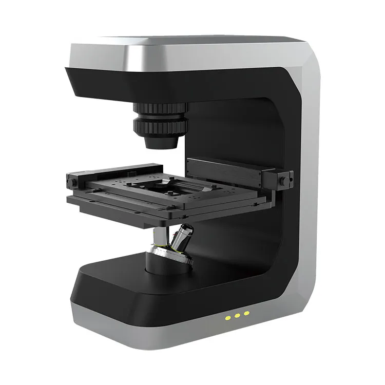
A 3D cell microscope is a specialized imaging tool used to capture and analyze three-dimensional images of cells and tissues. This type of microscope allows researchers and scientists to view cellular structures in three dimensions, providing more detailed and accurate information than traditional two-dimensional microscopy. Here’s an overview of 3D cell microscopes:
Key Features
1. Imaging Technology
- Confocal Microscopy: Uses a laser to scan and capture images at different depths within a sample, creating high-resolution 3D images by combining multiple 2D slices.
- Two-Photon Microscopy: Employs two-photon excitation to achieve deeper tissue penetration and reduced photodamage, suitable for imaging living cells and tissues.
- Light Sheet Microscopy: Illuminates the sample with a sheet of light and captures images from orthogonal angles, enabling fast and gentle 3D imaging with minimal phototoxicity.
2. Sample Preparation
- Fluorescent Labeling: Often involves labeling cellular components with fluorescent dyes or proteins to enhance visibility and contrast in 3D images.
- Staining and Fixation: Samples may be stained and fixed to preserve cellular structures and improve image quality.
3. Resolution and Depth
- High Resolution: Provides high spatial resolution to visualize fine details within cells and tissues.
- Depth Penetration: Capable of imaging through multiple layers of cells, offering a comprehensive view of cellular architecture.
4. Data Acquisition and Processing
- Image Stacking: Captures multiple images at different focal planes and stacks them to create a 3D representation.
- Software Analysis: Advanced software tools for processing, analyzing, and visualizing 3D images, including features like volume rendering, segmentation, and quantification.
5. Versatility
- Live Cell Imaging: Some systems are designed for live cell imaging, allowing researchers to observe dynamic processes in real-time.
- Fixed Samples: Suitable for analyzing fixed samples to study static cellular structures.
Advantages
1. Enhanced Visualization
- Three-Dimensional Detail: Provides a more complete and detailed view of cellular structures, allowing for better understanding of spatial relationships and organization.
- Depth Information: Offers insights into the depth and volume of cellular features, which is not possible with traditional 2D microscopy.
2. Improved Accuracy
- Structural Insights: Facilitates the study of complex cellular arrangements, interactions, and structures with greater accuracy.
- Quantitative Analysis: Enables precise measurement of cellular dimensions and volumes.
3. Dynamic Studies
- Live Imaging: Allows observation of dynamic cellular processes and interactions in living samples, providing valuable information about cellular behavior and function.
Applications
1. Cell Biology: Used to study cellular morphology, organelle structure, and cellular processes in both fixed and live cells.
2. Developmental Biology: Helps in understanding developmental processes and tissue organization during growth and differentiation.
3. Neuroscience: Facilitates the study of neuronal networks, synaptic connections, and brain structure.
4. Cancer Research: Provides insights into tumor cell architecture, metastasis, and the effects of therapeutic interventions.
Considerations
1. Sample Preparation: Proper preparation and staining of samples are crucial for obtaining high-quality 3D images.
2. Resolution vs. Depth: Balancing resolution and imaging depth can be challenging; higher resolution often comes with reduced depth penetration.
3. Data Management: 3D imaging generates large volumes of data, requiring robust data storage and processing capabilities.
Summary
A 3D cell microscope is a powerful tool for obtaining detailed and accurate three-dimensional images of cells and tissues. By using advanced imaging technologies such as confocal, two-photon, and light sheet microscopy, these systems provide enhanced visualization of cellular structures and dynamic processes. The ability to capture and analyze 3D images allows researchers to gain deeper insights into cellular biology and tissue organization, making it an invaluable tool in various fields of biological and medical research.